Leishmaniasis encompasses a spectrum of clinical manifestations caused by infection with the intracellular protozoan parasite Leishmania. [1] Transmission occurs through the bite of an infected female phlebotomine sandfly. [2, 3]
The most common presentation is cutaneous leishmaniasis, characterized by painless, chronic skin lesions ranging from nodules to large ulcers. These lesions typically persist for months to years but eventually resolve, often leaving scars.
Mucosal leishmaniasis, a less frequent but more severe form, affects the mucous membranes of the nose, mouth, and throat. It can lead to significant tissue destruction and disfigurement, causing severe functional and cosmetic impairment.
Visceral leishmaniasis, the most severe form, involves systemic dissemination of the parasite. Clinical features include irregular fever, hepatosplenomegaly, pancytopenia, and polyclonal hypergammaglobulinemia. Untreated visceral leishmaniasis carries a high mortality rate.
Treatment for leishmaniasis depends on the specific form and severity of the disease. However, therapeutic options are often limited, and drug resistance poses a growing challenge in disease management. With the exception of Australia, the Pacific Islands, and Antarctica, the parasites have been identified throughout large portions of the world.
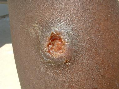
Classic Leishmania major lesion from a case in Iraq shows a volcanic appearance with rolled edges.
Classification
The taxonomy of Leishmania organisms is complex, and no single categorization is generally accepted. The 2 simplest and most widely used systems for categorizing leishmaniasis are as follows:
-
Categorization by clinical disease: In this system, leishmaniasis is divided into 3 primary clinical forms: cutaneous (localized, diffuse (disseminated), leishmaniasis recidivans, post–kala-azar dermal leishmaniasis), mucocutaneous , and visceral (kala-azar or black-fever in hindi ) and viscerotropic
-
Categorization by geographic occurrence: In this system, disease is divided into (1) Old World leishmaniasis (caused by Leishmania species found in Africa, Asia, the Middle East, the Mediterranean, and India), which produces cutaneous or visceral disease, and (2) New World leishmaniasis (caused by Leishmania species found in Central and South America), which produces cutaneous, mucocutaneous, and visceral disease
Signs and symptoms
Cutaneous leishmaniasis includes the following features:
-
Localized cutaneous leishmaniasis: Crusted papules or ulcers on exposed skin; lesions may be associated with sporotrichotic spread
-
Diffuse (disseminated) cutaneous leishmaniasis: Multiple, widespread nontender, nonulcerating cutaneous papules and nodules; analogous to lepromatous leprosy lesions
-
Leishmaniasis recidivans: Presents as a recurrence of lesions at the site of apparently healed disease years after the original infection, typically on the face and often involving the cheek; manifests as an enlarging papule, plaque, or coalescence of papules that heals with central scarring (ie, lesions in the center or periphery of an old healed leishmaniasis scar); relentless expansion at the periphery may cause significant facial destruction similar to the lupus vulgaris variant of cutaneous tuberculosis
-
Post–kala-azar dermal leishmaniasis: Develops months to years after the patient's recovery from visceral leishmaniasis, with cutaneous lesions ranging from hypopigmented macules to erythematous papules and from nodules to plaques; the lesions may be numerous and persist for decades
Mucocutaneous leishmaniasis consists of the relentless destruction of the oropharynx and nose, resulting in extensive midfacial destruction. Specific signs and symptoms include the following:
-
Excessive tissue obstructing the nares, septal granulation, and perforation; nose cartilage may be involved, giving rise to external changes known as parrot's beak or camel's nose
-
Possible presence of granulation, erosion, and ulceration of the palate, uvula, lips, pharynx, and larynx, with sparing of the bony structures; hoarseness may be a sign of laryngeal involvement
-
Gingivitis, periodontitis
-
Localized lymphadenopathy
-
Optical and genital mucosal involvement in severe cases
Visceral and viscerotropic leishmaniasis include the following features:
-
Visceral leishmaniasis (kala-azar): Potentially lethal widespread systemic disease characterized by darkening of the skin as well as the pentad of fever, weight loss, hepatosplenomegaly, pancytopenia, and hypergammaglobulinemia
-
Viscerotropic leishmaniasis: Nonspecific abdominal tenderness, fever, rigors, fatigue, malaise, nonproductive cough, intermittent diarrhea, headache, arthralgias, myalgias, nausea, adenopathy, and transient hepatosplenomegaly
Diagnosis
Laboratory diagnosis of leishmaniasis can include the following:
-
Isolation, visualization, and culturing of the parasite from infected tissue
-
Serologic detection of antibodies to recombinant K39 antigen
-
Polymerase chain reaction (PCR) assay for sensitive, rapid diagnosis of Leishmania species
Other tests that may be considered include the following:
-
CBC count, coagulation studies, liver function tests, peripheral blood smear
-
Measurements of lipase, amylase, gamma globulin, and albumin
-
Leishmanin (Montenegro) skin testing (LST) (not FDA approved in the United States)
Management
Treatment is tailored to the individual, because leishmaniasis is caused by many species or subspecies of Leishmania.
Pharmacologic therapies include the following:
-
Pentavalent antimony (sodium stibogluconate or meglumine antimonate): Used in cutaneous leishmaniasis; not marketed in the United States, but available through the CDC under an Investigational New Drug (IND) protocol
-
Liposomal amphotericin B (AmBisome): Effective against pentavalent antimony ̶ resistant mucocutaneous disease and visceral leishmaniasis
-
Oral miltefosine (Impavido): Approved by the FDA in March 2014 for visceral leishmaniasis due to L donovani; cutaneous leishmaniasis due to L braziliensis, L guyanensis, and L panamensis; and mucosal leishmaniasis due to L braziliensis
-
Intramuscular pentamidine: Effective against visceral leishmaniasis but associated with persistent diabetes mellitus and disease recurrence
-
Orally administered ketoconazole, itraconazole, fluconazole, allopurinol, and dapsone: None is as effective as the pentavalent antimony compounds, but they may be useful in accelerating cure in patients with cutaneous leishmaniasis that does not progress to mucosal disease and tends to self-resolve
-
Topical paromomycin: Shown to be effective against cutaneous leishmaniasis caused by L major and L mexicana
-
Sitamaquine: Undergoing phase 3 trials
Local therapies for some forms of cutaneous leishmaniasis include the following:
-
-
Local heat therapy at 40-42°C
Other important issues in the management of leishmaniasis are as follows:
-
Correction of malnutrition
-
Treatment of concurrent systemic illness (eg, HIV disease or tuberculosis)
-
Control of local infection